Your Patellofemoral Ligaments and Cartilage
Your patellofemoral knee joint is a complex and interconnected system of bones, ligaments, tendons, and cartilage. While this joint refers to the small area of interaction between your patella and femur, it is involved in many daily activities. Things like bending your knee and going up and down the stairs rely on smooth and controlled movements originating from your patellofemoral joint.
In this article, we go over the two main structures that stabilize and promote friction-free movement within your patellofemoral knee joint. These structures are the ligaments and articular cartilage. Before diving into these structures, this article also provides a brief overview of the structures that create movement in the patellofemoral joint.
How Knee Bends Happen: A Brief Overview
Knee bends are possible thanks to several key structures in your patellofemoral joint. Also called knee flexion, bending your knee happens as a result of the teamwork between your quadriceps muscle, patella, and quadriceps and patellar tendons.
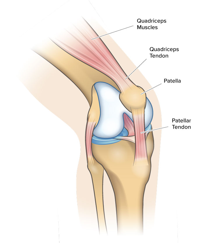
Together, these structures work like a pulley system. Your quadriceps and patellar tendons are like ropes that loop around your patella. When you activate your quadriceps muscle, force travels through your quadriceps tendon, patella, and patellar tendon to your shin bone (tibia). Because it can move between the two tendons, your patella ensures your knee movements are as efficient as possible.
If you are experiencing pain or difficulty performing leg movements, you may have damage in one of these structures. For a more in-depth understanding of the structures involved in knee bending, go to Patellofemoral Joint Explained.
Patellofemoral Ligaments vs. Tendons
While tendons promote movement at your joints, ligaments stabilize your joints by restricting motion. As discussed above, the quadriceps tendon and patellar tendon work together to make knee flexion possible.
Tendons and ligaments are similar because they are both tough, fibrous tissues that connect structures together. However, tendons typically connect bone to muscle while ligaments connect bone to bone. Some tendons, like the patellar tendon, connect bone to bone as a ligament would. But in these cases, these tissue bundles are called tendons rather than ligaments because their main function is helping the joint move.
Patellofemoral Ligaments Explained
Smooth and controlled knee flexion relies on ligaments that hold the patella in place. Like anchors, these fibrous tissues prevent movement by attaching structures to one another.
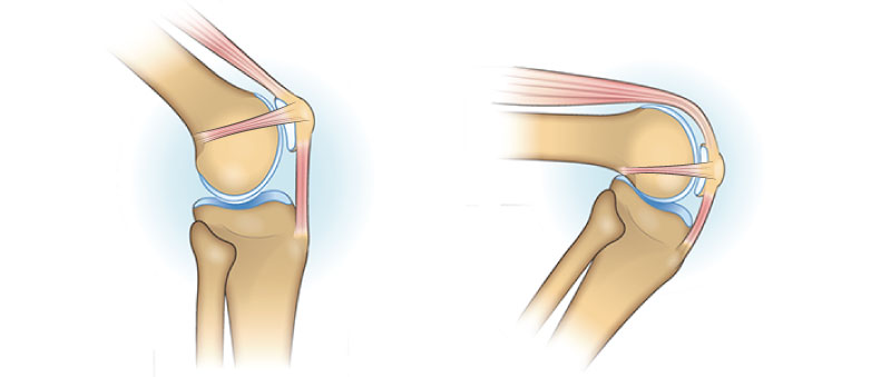
There are multiple ligaments that stabilize your patella. On the inner (medial) side of your knee, researchers have identified four different patellar ligaments1. The ligaments on the other (lateral) side of your knee are slightly different. However, both sides of your knee have patellofemoral and patellotibial ligaments. These ligaments connect the patella to the femur and tibia, respectively.
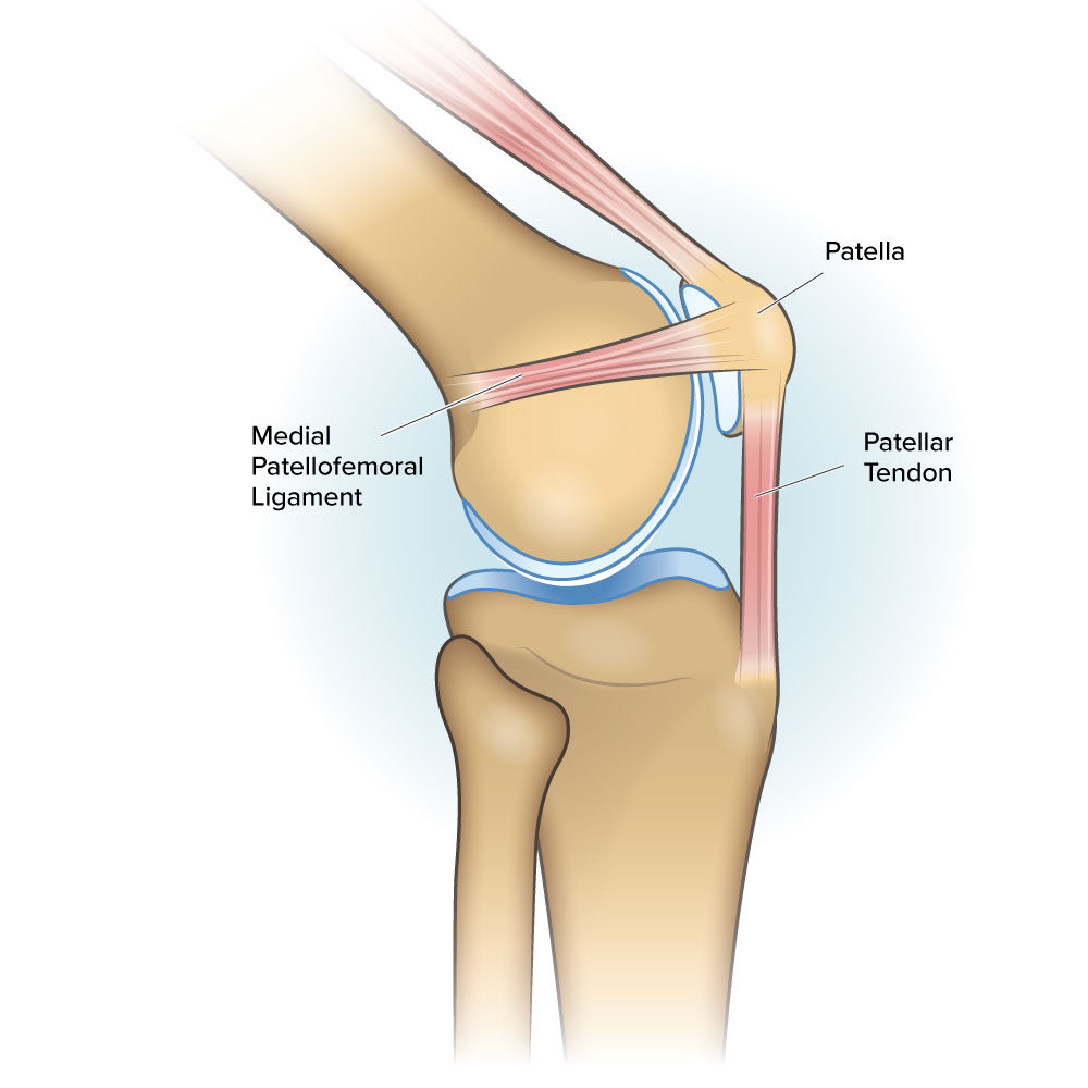
Based on how the ligament is named, you will know which structures are being connected. For example, the medial patellotibial ligament anchors the medial side of the patella to the tibia. The lateral patellotibial ligament connects the lateral side of the patella to the tibia.
Ligaments that are bundled with other ligaments are known as complexes. For example, the ligaments that attach your medial patella to your femur are known as the medial patellofemoral complex2.
Researchers are still working on understanding the specific ligaments that anchor down the patella. The purpose of these further studies is to help us understand how patellofemoral knee injuries happen.
Patellofemoral Ligament Injuries
Damage to your patellofemoral ligaments can affect the stability of your knee. Ligament injuries include sprains, tears, and complete ruptures. These can occur as a result of a forceful impact or twist to the knee.
Sprains occur when the ligament gets overstretched and damaged as a result. A similar word, strain, refers to overstretching of a muscle or tendon. Ligament tears, on the other hand, refer to breakages of the tissue. Tears can be categorized into three types – grades 1, 2, or 3 – based on their severity.
One of the most common tears is an ACL tear. This type of injury occurs when the anterior cruciate ligament, which attaches the tibia to the femur, gets damaged. ACL tears commonly occur when the leg is twisted while the foot is planted on the ground. For example, if you were doing a quick change of direction during a soccer game.
- For more information about ACL injury, take a look at the ACL Injury Patient Resource Guideline.
However, the ACL is not considered a structure of the patellofemoral joint, since it does not attach to the patella. While less common than tearing your ACL, it is possible to injure the ligaments that attach the patella to the other bones of the leg. These injuries are especially common if your knee gets dislocated.
Patellofemoral knee ligament tears, specifically in the medial patellofemoral complex, occur in approximately 95% of first-time knee dislocations3. Injury to patellar ligaments often leads to patellar instability and recurrent knee dislocation4.
Iliotibial Band Syndrome
Iliotibial Band Syndrome, or ITB syndrome, occurs when your iliotibial band is overused and irritated. Your ilitiobial (IT) band is a long piece of connective tissue that runs along your outer leg to connect your pelvis to your tibia. Although ITB syndrome does not originate in the knee, it can still cause patellar pain and knee instability.
The reason ITB syndrome can be felt in the knee is because of the lateral patellofemoral ligament. This ligament, found on the lateral side of your knee, connects your patella to your femur via the iliotibial band.
ITB syndrome is a type of overuse injury. Also called Runner’s Knee, this condition is caused by repetitive motions, such as running. While pain from ITB syndrome can be felt in the patellofemoral compartment, it can also be felt on the outside of your knee. Read about the five reasons for knee pain on the outside of your knee here.
Articular Cartilage of the Patellofemoral Knee Joint
Your knee is classified as a synovial joint, which means that the place where your bones contact each other is enclosed in a membrane called a “joint cavity.” The bones that make up this cavity have a layer of what’s called articular cartilage on their surface.

Articular cartilage is a type of white, connective tissue that lines your bones in areas where they interact with other bones. It does not contain blood vessels or nerves, and if damaged, has a limited ability to heal itself.
The joint cavity also contains synovial fluid. The purpose of articular cartilage and synovial fluid is to reduce friction between your bones and help distribute and absorb the forces passing through your joint.
Within your patellofemoral knee joint, you will find articular cartilage on the underside of your patella and within the trochlear groove of your femur. As you flex and extend your knee, your patella moves up and down the trochlear groove. This movement is called patellar tracking. The articular cartilage and synovial fluid within the joint cavity help ensure that this motion is smooth and painless.
Damage to Your Articular Cartilage
Damage to the articular cartilage damage on your patella and trochlear groove can be painful and debilitating. Articular cartilage loss can be caused by:
Wear and Tear: As you and your knees age, you may experience the natural wearing down of your cartilage. This type of articular cartilage damage is called patellofemoral arthritis.
Traumatic injury: If you put a large and unexpected amount of force through your knee, your cartilage can become damaged. This can occur from a bad fall, a car accident, or high-impact sports. Cartilage damaged from a traumatic injury can result in early-onset arthritis.
Lack of movement: It should be no surprise that you need to keep your body moving to remain healthy. Articular cartilage does not have a blood supply, so it relies on the synovial fluid within your joint cavity to deliver the oxygen and nutrients it requires to remain healthy. Synovial fluid is thick and needs movement to help it flow. If you have a lack of movement at your knee joint, your articular cartilage may become deprived of these nutrients.
References
- B. M. Kruckeberg et al., “Quantitative and Qualitative Analysis of the Medial Patellar Ligaments: An Anatomic and Radiographic Study,” Am J Sports Med, vol. 46, no. 1, pp. 153–162, Jan. 2018, doi: 10.1177/0363546517729818.
- A. E. Loeb and M. J. Tanaka, “The medial patellofemoral complex,” Curr Rev Musculoskelet Med, vol. 11, no. 2, pp. 201–208, Jun. 2018, doi: 10.1007/s12178-018-9475-2.
- A. S. Panni, M. Vasso, and S. Cerciello, “Acute patellar dislocation. What to do?,” Knee Surg Sports Traumatol Arthrosc, vol. 21, no. 2, pp. 275–278, Feb. 2013, doi: 10.1007/s00167-012-2347-1.
- C. Krebs, M. Tranovich, K. Andrews, and N. Ebraheim, “The medial patellofemoral ligament: Review of the literature,” J Orthop, vol. 15, no. 2, pp. 596–599, May 2018, doi: 10.1016/j.jor.2018.05.004.