Your Patellofemoral Knee Joint Explained
Your patellofemoral knee joint plays an important role in your everyday life. This joint is crucial for movements such as climbing stairs, walking up a slope, running, standing from seating, and many other daily tasks.
We don’t often think about our joints and how they work until something goes wrong. If you are experiencing patellofemoral knee pain (pain originating under the kneecap), you may be wondering: What is happening inside my knee that is causing me so much grief?
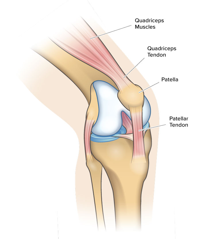
To give you a better understanding, this article will go over three major patellofemoral knee joint structures that can contribute to knee pain. These major structures include the quadriceps muscles, quadriceps tendon, and patellar tendon.
Your Knee Joint
A joint is classified as a point in the body where two bones meet. Believe it or not, your body has approximately 300 joints! The joints of the human body are categorized depending on how they move.
Your patellofemoral knee joint, along with your elbow, fingers, and toes, are called hinge joints. Like a door hinge, hinge joints only move in one direction. Don’t let the simplicity of a hinge joint motion fool you. The knee is one of the largest and most complex joints of the body.
To understand how a hinge joint works, try moving your hip and then move your knee. You should notice that your hip joint has the ability to move your leg forwards, sideways, and in a circular motion. In comparison, your knee joint can only move your lower leg forward and backward.
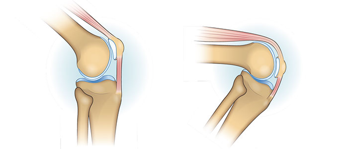
These two movements of your knee joint are called knee extension and knee flexion. Knee extension is when you are straightening your knee. Knee flexion is when you are bending your knee.
Bones of Your Knee
Your knee joint involves three bones. Two of these bones – your femur (thigh bone) and your tibia (shin bone) – are known as long bones. The purpose of long bones is to support your body and keep it upright.
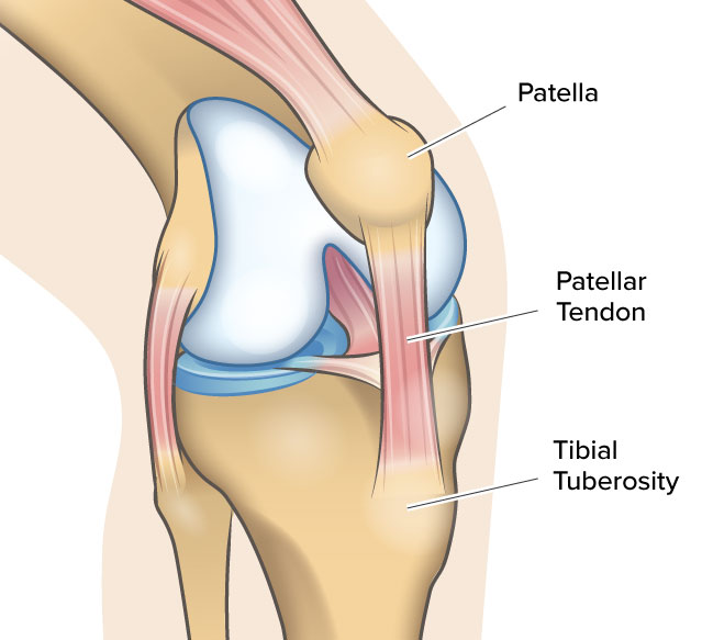
The third type of bone is your patella (commonly referred to as your kneecap), which is a sesamoid bone. Sesamoid bones are embedded in tendons. The patella is located between your patellar tendon and quadriceps tendon.
Knee Compartments
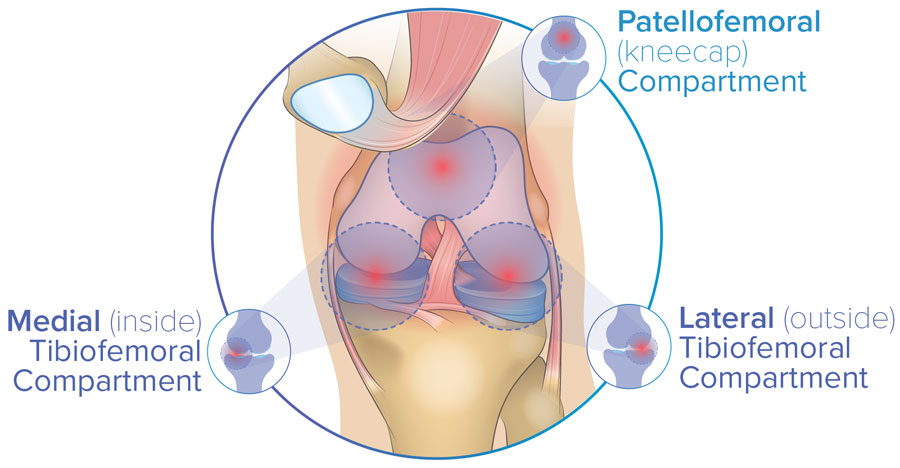
The femur, tibia, and patella create three separate knee joint compartments. The tibia and femur meet to form two of these compartments on the inside and outside of your knee joint. These compartments are called the medial and lateral tibiofemoral compartments.
The third compartment occurs where your patella moves along the groove in your femur. This groove is called the trochlear groove and the overall area of this interaction is called the patellofemoral compartment.
In addition to your bones, there are many structures that work together to create the knee joint. These other structures include your meniscus, articular cartilage, tendons, and muscles. Each of these structures serves a unique purpose. Unfortunately, each of these structures can also be damaged, leading to many different types of knee injuries.
To see how all these structures fit together, click here for a video on knee anatomy.
The Purpose of Your Patellofemoral Knee Joint
Have you ever wondered why you have a kneecap? The most obvious reason is for protection. If you fall directly on your knees, the hard bony surface of your patella will protect the delicate structures within your knee from trauma. In addition to acting as a shield, the patella also protects the patellar and quadriceps tendon from frictional forces1. Although protection is an important function of the patella, it’s not the only function.
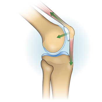
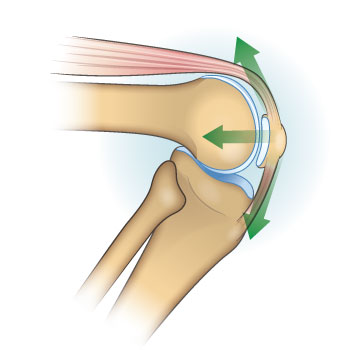
One of the most important functions of your patella is to make it easier to extend your knee. In this case, your patella works like a pulley. Imagine your patellar tendon and quadriceps tendons as ropes that loop over your patella. By looping around your patella, these tendons can redistribute force across your leg much more easily.
With the pulley system created by your patella, knee extension works as follows. Your quadriceps muscles create a force that travels across your quadriceps tendon, patella, and patellar tendon. This force travels across these structures until it meets the tibia. Finally, knee extension occurs when the force pulls on your tibia.
So how important is your patella in knee extension? Your patella increases the efficiency of your quadriceps muscles by approximately 60%2. Your knee is very impressive! To top it off, the way your patella moves within its trochlear groove ensures that knee extension is as efficient as possible3.
Muscles & Tendons of Your Patellofemoral Joint
Knee extension is possible thanks to the teamwork between your muscles, tendons, and ligaments in your patellofemoral knee joint. There are many other structures in your patellofemoral knee joint. However, you can gain a fairly good understanding of the patellofemoral knee joint by first learning about three related structures. These three structures are the quadriceps muscle, quadriceps tendon, and patellar tendon.
Quadriceps
One of the most powerful groups of muscles in your body is the quadriceps muscle. Located on the front of your upper leg, the quadriceps controls the motion of your patellofemoral knee joint.
The quadriceps is actually a collection of four individual muscles. These muscles include the rectus femoris, vastus lateralis, vastus medialis, and vastus intermedium.
Quadriceps Tendon
Tendons and ligaments are tough fibrous tissues that connect structures together. Tendons connect bone to muscle with the purpose of moving the joint. In comparison, ligaments connect bone to bone and provide stability.
Your quadriceps tendon is made from the four quadriceps muscles that merge together. It connects your quadriceps muscles to the upper part of your patella.
During knee extension, your quadricep muscles and tendon apply a pulling force on your patella. As your bend your knee, these forces become stronger. When climbing stairs, for example, these forces apply a load on your knee that is equivalent to 3.3 times your body weight4.
Patellar Tendon
Your patella is suspended between two tendons. While the tendon above the patella is the quadriceps tendon, the tendon below is called the patellar tendon. The patellar tendon connects the lower part of your patella to a bump on the upper part of your tibia. That bump is called your tibial tuberosity.
Your patellar tendon helps you perform knee extension. Working with the quadriceps muscle, quadriceps tendon, and patella, the patellar tendon helps apply a force to the tibia.
A Note on the “Patellar Ligament”
If you recall our earlier definitions of ligaments and tendons, you may wonder why the patellar tendon is even called a tendon. To recap, ligaments connect bone to bone while tendons connect muscle and bone. However, the patellar tendon connects two bones: your patella and tibia. So, shouldn’t this structure be called the patellar ligament instead?
In this case, we look at the function of the structure rather than what it connects. The patellar tendon is called a tendon instead of a ligament because it works more like a tendon. To refresh your memory, tendons help move your joints while ligaments help stabilize them.
So even though the patellar tendon connects bone to bone, it helps move the joint rather than restrain it. As a result, this fibrous tissue is commonly referred to as a tendon. However, if you hear the term “patellar ligament,” know that it is referring to the same structure.
Muscle & Tendon Injuries of the Patellofemoral Joint
There are numerous patellofemoral injuries and diseases that can occur from muscle and tendon damage. A few examples include strains and tears, tendonitis, tendinosis, and quadriceps weakness.
Strains and Tears
Strains and tears can have similar symptoms. However, the severity of their damage often differs. A strain occurs when the muscle or tendon is overstretched. Tears are more severe than strains and involve the breakage of fibers in your muscles or tendons.
Tears are classed into three different categories depending on the severity. Grade 1 and 2 tears are partial tears. A partial tear occurs if some of the fibers within the muscle or tendon break. Think about a rope that gets stretched so far that it frays, but does not snap.
Grade 3 tears, also called complete ruptures, occur when the quadriceps muscle is completely separated from the tendon and patella. Since our bodies work hard every day, strains and minor tears are quite common. However, it is important to let your body heal so that your injury does not become worse.
Also, try not to confuse strains with a similar word, sprain. A sprain refers to the overstretching and damage of ligaments. This overstretching can occur for a variety of reasons. Some of these reasons include not properly warming up, exercising when you are fatigued or when your muscles are weak. Sprains can also happen if you stretch an area suddenly or unexpectedly. For example, catching yourself during a fall can result in a sprain.
Tendonitis or Tendinosis
If you are have a tendon injury, you will need to be patient. Tendons heal slowly because they do not have a large blood supply. Two types of tendon injuries include tendonitis and tendinosis.
Tendonitis occurs when your tendon becomes painful and inflamed due to the accumulation of very small tears. This is an overuse injury, which means that it can occur if you perform the same movement many times. Quadriceps tendonitis is common in runners and people who play sports requiring jumping.
In comparison, tendinosis is a chronic condition that involves the degeneration of the tendon tissue. It also creates pain and swelling in your knee. But instead of tiny tears, tendinosis involves the fibers of your tendon becoming thick, hard, and tangled.
Tendinosis can occur if the tendon is constantly damaged and isn’t given enough time to heal. The condition is commonly misdiagnosed as tendinitis. However, ultrasound imaging can help tell these conditions apart5.
Quadriceps Weakness
Strong quadriceps are required to support your body weight, extend your leg, and stabilize your patella. When your quadriceps become weak, you can find these movements more challenging and painful.
Quadriceps weakness can occur when the muscle is not being used. However, it may also occur from an injury or disease. Medical conditions that cause quadriceps weakness include stroke, muscle dystrophy, and multiple sclerosis. These diseases either affect the nerves that control your muscles or your body’s ability to build and sustain muscle.
In the case of your quadriceps muscles, the phrase “use it or lose it” is applicable. For example, two weeks of complete bed rest can cut the mass of your quadriceps muscles in half6.
Protecting the Muscles & Tendons of Your Patellofemoral Knee Joint
Your patellofemoral knee joint is a complex system with many interrelated structures. As a result, many things can go wrong in your patellofemoral knee joint.
Research suggests that approximately 25% of the population experiences patellofemoral knee pain7. Unfortunately, the long-term outcomes for many of these conditions are not reversible. Therefore, it is crucial to understand what actions you can take to help avoid the diseases that cause patellofemoral knee pain.
Take Precautions During High-Risk Activities
Activities with repeated motions or with a high likelihood of traumatic impact to your knee may increase the risk of developing patellofemoral knee pain. Some of these high-risk activities include running and jumping, football, soccer, and military training.
If you decide to partake in these activities, ensure you are using the right equipment. You can also consider switching up your training with low-impact activities. Sports like swimming and biking can help give your patellofemoral knee joint a rest.
Maintain A Healthy Weight
Every time you take a step, the weight of your body travels down through your knee joints. If you are carrying extra body weight, you are requiring your knees to undergo higher joint forces. Studies show that a higher BMI can lead to a higher risk of knee conditions such as osteoarthritis8.
As a result, maintaining a healthy BMI can help prevent patellofemoral knee pain. Weight management is considered one of the top conservative treatment methods for managing knee pain.
Keep Your Quadriceps Muscles Healthy
Strong quadriceps muscles are a key component of patellar stability. You need healthy quadriceps to help control your knee movements. They also help prevent knee pain and injury by absorbing forces while you go about your daily activities.
To keep your quadriceps healthy, ensure you eat a proper diet with enough protein. It is also beneficial to incorporate lower-body exercises and stretches into your routine.
Wear A Patellofemoral Knee Brace
If you require extra stability, are struggling with weight loss, have quadriceps weakness, or require offloading of your patellofemoral joint, bracing is a great conservative option for you to manage your symptoms. Knee braces can help delay or remove the need for patellofemoral knee surgeries, or aid in your recovery post-surgery.
The Spring Loaded Tri-Compartment Offloader
The Spring Loaded Tri-Compartment Offloader knee brace is the only brace on the market with clinically proven knee offloading in all three compartments of the knee, including the patellofemoral compartment9,10.
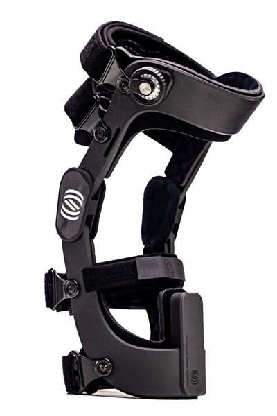
Read more about the Spring Loaded brace and other patellofemoral knee braces here.
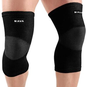
Compression Knee Braces
For more minor conditions of the patellofemoral knee joint, you can also consider wearing a compression knee brace. Mild conditions suitable for this type of bracing include patellar misalignment or mild tendon swelling. These braces may also help prevent further wear and tear of your patellofemoral compartment.
References
- C. F. Cox, M. A. Sinkler, and J. B. Hubbard, “Anatomy, Bony Pelvis and Lower Limb, Knee Patella,” in StatPearls, Treasure Island (FL): StatPearls Publishing, 2021. Accessed: Jun. 20, 2021. [Online]. Available: http://www.ncbi.nlm.nih.gov/books/NBK519534/
- F. Lieb and J. Perry, “Quadriceps Function: An Anatomical And Mechanical Study Using Amputated Limbs,” The Journal of Bone & Joint Surgery, vol. 50, no. 8, pp. 1535–1548, Dec. 1968.
- A. Fox, F. Wanivenhaus, and S. Rodeo, “The Basic Science of the Patella: Structure, Composition, and Function,” J Knee Surg, vol. 25, no. 02, pp. 127–142, Jun. 2012, doi: 10.1055/s-0032-1313741.
- D. T. Reilly and M. Martens, “Experimental Analysis of the Quadriceps Muscle Force and Patello-Femoral Joint Reaction Force for Various Activities,” Acta Orthopaedica Scandinavica, vol. 43, no. 2, pp. 126–137, Jan. 1972, doi: 10.3109/17453677208991251.
- E. Bass, LMT, “Tendinopathy: Why the Difference Between Tendinitis and Tendinosis Matters,” IJTMB, vol. 5, no. 1, pp. 14–17, Mar. 2012, doi: 10.3822/ijtmb.v5i1.153.
- A. Shaibani, Quadriceps Weakness, vol. 1. Oxford University Press, 2015. doi: 10.1093/med/9780199898152.003.0014.
- B. E. Smith et al., “Incidence and prevalence of patellofemoral pain: A systematic review and meta-analysis,” PLoS ONE, vol. 13, no. 1, p. e0190892, Jan. 2018, doi: 10.1371/journal.pone.0190892.
- Z.-Y. Zhou, Y.-K. Liu, H.-L. Chen, and F. Liu, “Body mass index and knee osteoarthritis risk: A dose-response meta-analysis: Body Mass Index and Knee Osteoarthritis Risk,” Obesity, vol. 22, no. 10, pp. 2180–2185, Oct. 2014, doi: 10.1002/oby.20835.
- A. R. Budarick, B. E. MacKeil, S. Fitzgerald, and C. D. Cowper-Smith, “Design Evaluation of a Novel Multicompartment Offloader Knee Brace,” J Biomech Eng, vol. 142, no. 1, Jan. 2020, doi: 10.1115/1.4044818.
- C. A. McGibbon, S. Brandon, E. L. Bishop, C. Cowper-Smith, and E. N. Biden, “Biomechanical Study of a Tricompartmental Offloader Brace for Patellofemoral or Multicompartment Knee Osteoarthritis,” Front. Bioeng. Biotechnol., vol. 8, 2021, doi: 10.3389/fbioe.2020.604860.